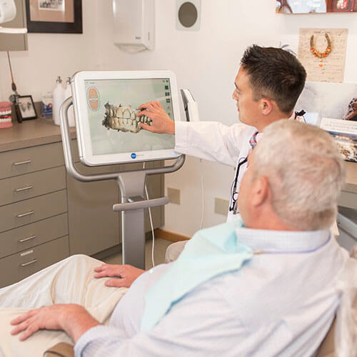
Routine x-rays can be a conflicting topic in the dental office, especially while many people do not understand the benefits and importance of updating images. In some cases the answer could be getting images done every 18-24 months.

Those cases are rare and not usually advised, especially as we age. You wouldn’t want to have a large cavity that now needs an extraction that could have been avoided and fixed with a simple filling.
The need for radiographs is in part, based on your risk for developing decay, your habits, previous history of decay and many other factors. It is our job as dental professionals to tailor the need for radiographs to each patient.
That being said, a majority of individuals are at a moderate-high risk for developing decay, which is why in most cases yearly radiographs are highly recommended. Most insurances also follow these guidelines and typically cover x-rays as advised by your dentist(yearly).
Radiographs or X-Rays
Radiographs or x-rays provide the dentist with needed information that cannot be collected with a simple oral examination.
More often than not cavities that appear in the visual exam can only be seen on certain surfaces or teeth, or when they’ve already increased in size. Our objective is to always catch decay as soon as possible.
When we treat decay early on we can prevent extensive loss of your tooth structure and if we do have to take excessive amounts it can weaken your tooth.

By getting in early you are also avoiding the costs of more invasive treatment that would be needed otherwise. Most people would rather have a simple filling to treat early decay than a root canal and crown (or losing the tooth) because the decay has progressed.
Depending on your personal risk factors for decay, cavities can sometimes progress very rapidly, and sometimes changing very drastically within just 12 months, sometimes less.
Detailed Inside Look
X-rays give us a detailed inside look between each tooth you have, again an area that cannot be seen with just an oral exam. They also allow us to see the three layers of tooth structure (enamel, dentin, and pulp chamber) to determine what stage the cavity has advanced into.
When a cavity is still in the enamel layer, it will progress more slowly. When it surpasses the enamel and reaches the dentin layer of the tooth, it will progress more quickly. The accelerated advancement at this stage is due to the softer structure of the dentin.
Even if a cavity does not infect the pulp chamber (the inner portion of the tooth containing nerves and blood vessels), a cavity in that close of proximity, is sometimes just enough to disturb the nerve of the tooth and cause it to become inflamed, causing irreversible pulpitis, typically resulting in a root canal and worse case extraction.

Save Money, Time, and Pain
Allowing your doctor or dental assistant to perform the radiographs they deem appropriate can more than often save you time, money and pain by finding cavities while they are less advanced.
If you have any concerns, please do not hesitate to discuss them with your hygienist as well. It is never our goal to expose anyone to unnecessary radiation. In fact, all of our x-ray machines are completely digital (drastically decreasing radiation exposure) and lead aprons are used on everyone.
Dental x-rays have decreased dramatically in radiation over the years. We will discuss your particular risk factors and our recommendations for radiographs with you in office.
You are ultimately in control of your health care and have every right to determine what treatment you do or do not want to receive and we are willing to discuss this further at any of your future visits.

At Eleven Eleven Dental, we believe your comfort should always come first. You will feel our warmth and compassion as soon as you walk through our doors.